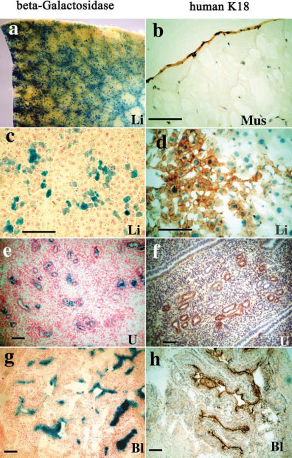
FIG. 6.

Immunohistochemical localization of K18 and β-gal activity of OHT-treated K18iresCreER;R26R mice. a: Whole-mount β-gal activity of liver (blue). b–h: Frozen sections of mouse tissues were stained for human K18 (b,d,f,h) or Cre-mediated β-gal activity (c,e,g). b: Localization of K18 in the epithelial covering of muscle. c,d: Hepatocytes stained for β-gal and K18 (brown). e,f: Uterine gland staining of the uterus. g,h: Transitional epithelium staining of the urinary bladder. Li, liver; Mus, muscle; U, uterus; Bl, bladder. Scale bars = 100 μm.
