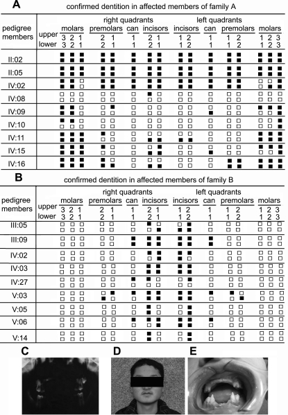
Figure 2. Clinical evaluations.
(A) and (B). Synopsis of the permanent dentition in affected members of Family A and Family B. Closed squares represent absent teeth. (C). Panoramic radiograph of a proband (IV:02) in Family A at age 14. He had up left permanent second molar, four permanent first molars and four milk second molars. (D) and (E): Clinical appearance of V:03 (Family B), at age 18, showing absence of all the incisors, two up cans and four premolars.

