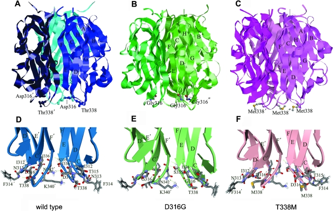
Figure 6. Structures of wild type, D316G and T338M EDA.
(A)–(C). Locations of p.D316G or p.T338M mutation sites in the quaternary structure of the EDA homotrimers. The EDA trimers are shown as ribbon rendering with mutation residues rendered in stick and ball, beta strands and the mutated amino acids are labeled. (D)–(F). Close up views of the mutation sites of D316G, T338M and wild type EDA proteins. The amino acid backbones are represented by ribbons with arrows. Side chains have been omitted from most residues for clarity. Hydrogen bonds are indicated by dashed green lines.

