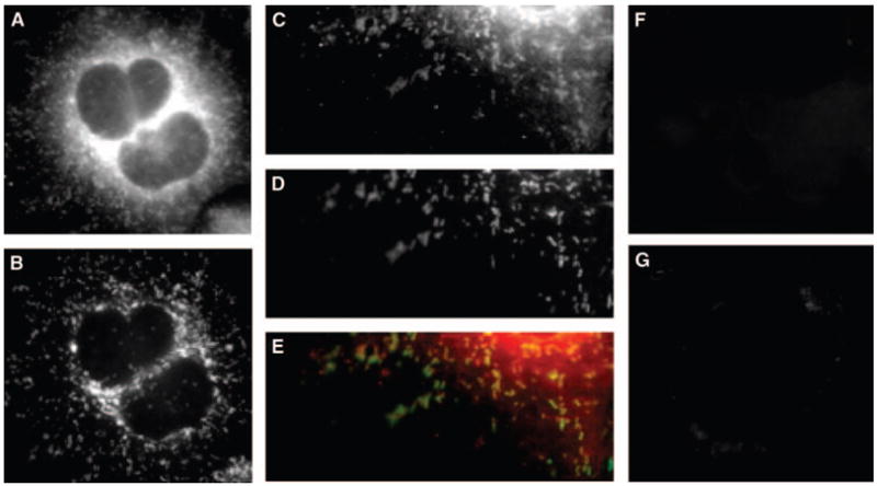
Fig. 7.

cPLA2γ localizes to mitochondria in IMLF−/−. IMLF−/− were infected with Ad-cPLA2γ, fixed, and probed with polyclonal anti-serum to anti-cPLA2γ and monoclonal antibodies to anti-oxidative phosphorylation complex V. Secondary antibodies used were conjugated to Texas Red and AlexaFluor 488, respectively. Immunofluorescence of cPLA2γ (A, C) and of the mitochondrial marker oxidative phosphorylation complex V (B, D) is shown. An overlay of cPLA2γ fluorescence (red) and mitochondrial marker fluorescence (green) is shown (E). IMLF−/− overexpressing cPLA2γ were probed with Texas Red secondary antibody only (F), and probed with anti-oxidative phosphorylation complex V primary monoclonal antibodies and anti-rabbit secondary Texas Red antibodies as controls (G).
