
Fig 2. Cdc42 siRNA-depletion delays lumen formation in MDCK cyst.
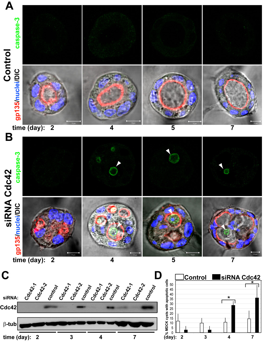
(A) and (B) Effect of Cdc42-1 siRNA on lumen formation and apoptosis. MDCK cells were transfected with Cdc42 siRNA (B) or siRNA control (A) and plated to form cysts. Cells were fixed at 2, 3, 4, 5 and 7 days and stained to detect gp135 (red) and nuclei (lower panels merged with DIC); and caspase3 (green, upper panels). Arrowheads indicate apoptotic cells in the lumen of the cysts. Scale bars 5 µm.
(C) Down-regulation of Cdc42 by siRNA. Cells were transfected with siRNAs Cdc42-1 and Cdc42-2 against canine Cdc42 or with control siRNA, allowed to form cysts for 2, 3, 4 or 7 days and then total cell lysates western blotted for Cdc42 and β-tubulin (control).
(D) Quantitation of cysts with apoptotic cells in the lumen in cells transfected with control siRNA (white bars), specific siRNA Cdc42-1 (black bars). Values shown are mean ±SD from 4 different experiments. * P≤0.01; P<0.01