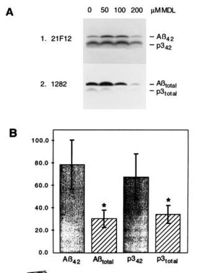
Figure 2.

Differential inhibition of Aβ42 and Aβ40 formation. (A) Labeled K695sw cells were chased with the indicated concentrations of MDL 28170 and precipitated with 21F12 (Upper) followed by 1282 (Lower). (B) Quantitation of the effect of 200 μM MDL 28170 on Aβ and p3 by phosphorimaging. The bars show the pixel number relative to an untreated control (200); SDs are indicated. The decreases in Aβtotal and p3total relative to an untreated control were significant (∗, two-tailed t test, n = 4, P < 0.001) and the decreases in Aβ42 and p342 upon treatment with MDL 28170 did not reach significance. Moreover, the difference in inhibition of Aβ42 versus total Aβ is significant (two-tailed t test, n = 4, P < 0.01) and the difference in inhibition of p342 versus total p3 is also significant (two-tailed t test, n = 4, P < 0.05).
