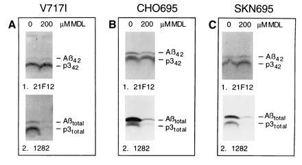
Figure 4.

Differential inhibition of Aβ42 and Aβ40 formation in different cell types. Labeled cells were chased with the indicated concentrations of MDL 28170, and the media were precipitated with 21F12 (Upper) followed by 1282 (Lower). (A) K695717I cells. The relatively low βAPP expression in this line leads to a faint Aβ42 band. However, the bands are not due to endogenous βAPP, because nontransfected 293 cells do not show any Aβ or p3 bands under the conditions of the experiment (not shown). (B) CHO695 cells. Note that in CHO cells, the p3 bands migrate as doublets, as described (30). (C) SKN695 cells.
