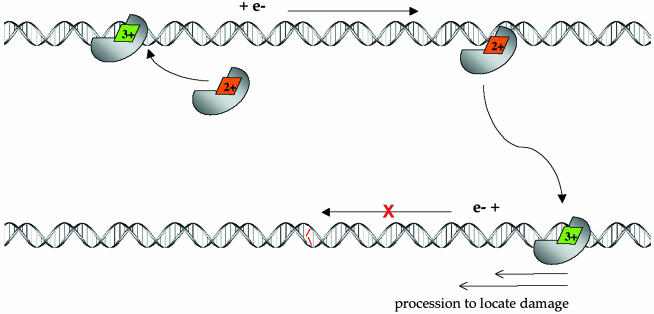
Fig. 5.
Schematic model of long-range scanning for mismatches by MutY through DNA-mediated CT chemistry. (Upper) Nonspecific binding of MutY to DNA, where binding is associated with a shift in redox potential of the [4Fe4S]2+ cluster, leading to oxidation to the 3+ form. Associated with cluster oxidation is DNA-mediated CT to an alternately bound MutY, where reduction to the 2+ cluster promotes dissociation of the protein. Because this CT process proceeds without interruption by an intervening mismatch, the process constitutes a scan of this region of the genome. (Lower) Association of MutY to a region containing a mismatch (red), where the associated stacking perturbation does not permit DNA-mediated CT to occur. Here, the protein is shown processively diffusing to the mismatch site.

