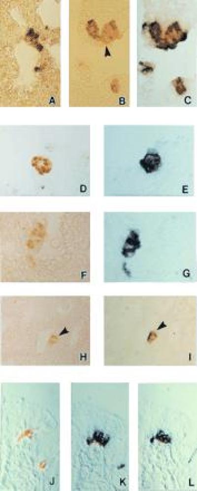
Figure 1.

NISH for gp91phox and p22phox subunits of the NADPH oxidase (O2-binding protein) components and KH2O2 channel KV3.3a in pulmonary NEB of 26 day gestation fetal rabbit lung. (A) NISH using the NADPH oxidase subunit gp91phox RNA antisense probe shows specific signal (purple) localized in NEB cells. The section was counter-stained with safranin O. (×400.) (B) 5-HT-immunoreactive NEB cells located within airway mucosa at bronchial bifurcation (arrowhead). (×400.) (C) NISH on the same section as in B using the NADPH oxidase subunit gp91phox RNA antisense probe shows specific mRNA signal (purple blue) localized in the same NEB cells. (×400.) (D) Immunostaining for 5-HT (light brown) combined with NISH using the NADPH oxidase subunit gp91phox RNA sense probe (negative control). There is slight focal background staining of lung interstitial cells but no mRNA signal is present in NEB cells indicating specificity of the procedure. (×400.) (E) Immunostaining for 5-HT followed by NISH with the NADPH oxidase subunit gp91phox RNA antisense probe on a serial section next to D. (×400.) (F and G) The same tissue and procedures as B and C using the NADPH oxidase subunit p22phox RNA antisense probe. Strong positive signal for p22phox mRNA (G) (purple color) is localized in 5-HT positive NEB cells (brown color) (F). Immunostaining for 5-HT followed by (H) NISH for KV3.3a using RNA antisense probe (I) showing specific mRNA signal (arrowhead) localized in the same 5-HT immunopositive NEB cells. (×250.) Immunostaining of NEB cells for 5-HT in H has been purposefully under developed, just enough to allow identification of NEB cells without possible interference of chromogen used in NISH. (J) Bombesin immunostaining on formalin fixed paraffin embedded sections of neonatal human lung followed by NISH using the KV3.3a sense RNA probe (negative control). Two bombesin immunoreactive foci (light brown) corresponding to NEB cells within airway epithelium. Absence of mRNA signal indicates specificity of the NISH reaction. (K) Bombesin immunostaining followed by NISH with the KV3.3a RNA antisense probe on a serial section next to J showing specific expression of the K+ channel mRNA in NEB cells. (L) Bombesin immunostaining followed by NISH with the NADPH subunit gp91phox RNA antisense probe on a serial section next to K demonstrating correlation with the K+ channel mRNA signal in the same NEB cells. (Nomarski interference contrast, ×400.)
