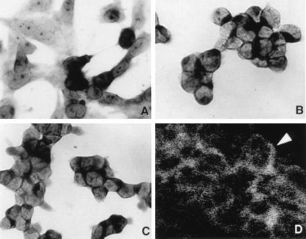
Figure 2.

NISH for gp91phox and KV3.3a with immunohistochemistry for p22phox in SCLC cell lines. (A) NISH for gp91phox using RNA antisense probe on H-69V line showing variable expression with strong positive signal in some cells and a weaker or no signal in adjacent tumor cells. (×400.) (B) NISH for gp91phox on NCI-H146 cell line showing more uniform cytoplasmic localization of mRNA signal. (C) NISH for KV3.3a using antisense RNA probe on NCI-H146 show similar signal distribution as in B. (×400.) (D) Immunohistochemistry for p22phox on NCI-H146 cell line with membranous or submembranous localization of positive immunoreactivity (arrowhead). (Laser confocal microscopy, fluorescein isothiocyantate-labeled secondary antibody, ×800.)
