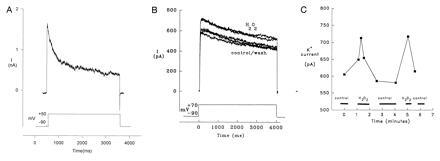
Figure 5.

Whole-cell currents in cultured fetal rabbit NEB cells. When NEB cells were exposed to a brief period of hyperpolarization followed by a longer depolarizing pulse, a slowly inactivating IA current was revealed (n = 15). (A) NEB cells shown was recorded in the presence of an extracellular bathing solution A and an intracellular pipette solution containing: 140 mM KCl, 1 mM CaCl2, 10 mM EGTA, and 10 mM Hepes. NEB cells were stepped from −90 mV to various depolarizing potentials for 3–4 sec (n = 8). The addition of 2 mM MgATP in the pipette solution did not alter the results. The slowly inactivating K+ current observed was similar to the one described in Xenopus oocytes KV3.3a channel subunit expressing cells (13). (B) An increase in the K+ current was observed when NEB cells were exposed to either brief or prolonged depolarizing pulses in the presence of varying concentrations of H2O2 (n = 3). The trace shown is of a NEB cell stepped from −90 mV to +70 mV for 4 sec in the presence of extracellular solution B. This solution was used to eliminate any contribution of Ca2+ and Na+ currents. There was a resultant increase in the K+ current in the presence of H2O2 (0.1–1.6 mM) that was reversible upon returning the cell to H2O2-free solution. This effect was repeated two times on the cell shown. (C) A graph depicting the change in maximal amplitude of the K+ current over time of the same cell as in B.
