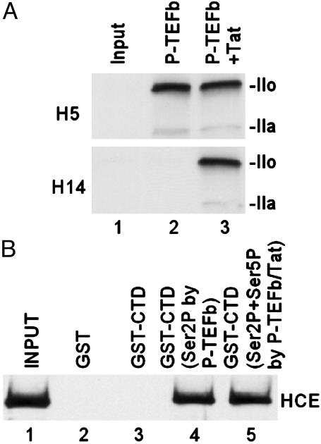
Fig. 1.
Interaction between HCE and CTD phosphorylated by P-TEFb. (A) Tat modifies the substrate specificity of CDK9. In vitro kinase assays were performed by incubating purified RNAP II with P-TEFb and ATP (100 μM) in the absence or presence of Tat. Phosphorylated CTD was analyzed by Western blot with specific antibody H5 (α-phosphoserine 2) or H14 (α-phosphoserine 5). The hypophosphorylated (IIa) and hyperphosphorylated (IIo) forms of the largest subunit of RNAP II are indicated. (B) Interaction of HCE and CTD phosphorylated by P-TEFb. HCE was incubated with bead-bound GST-CTD. Bead-bound proteins were eluted by boiling in SDS loading buffer and then analyzed by Western blot with α-CE antibody.

