
Figure 1.
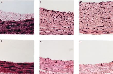
Histology of rat carotid arteries after balloon injury and various treatments as described below. All barriers were nondegradable and remained adherent to the artery wall for the duration of the experiment. The hydrogel barrier cannot be observed in these sections due to the dehydration process. Staining was by hematoxylin and eosin. Black arrows denote the location of the internal elastic lamina. (Bar = 50 μm.) (A) Control vessel 2 hr after injury. Platelet deposition can be clearly noted above the two arrowheads. (B) Hydrogel-treated vessel 2 hr after injury. No platelet deposition could be noted by light microscopy. (C) Control vessel 28 days after injury. The formation of a fibrocellular neointima is clearly evident above the two arrowheads. (D) Hydrogel-treated vessel 28 days after injury. Isolation from blood contact prevented neointima formation and slowed repopulation of the media. (E) Vessel 14 days after injury, with local delivery of PDGF but without blockade of blood contact. Under these conditions, local delivery of PDGF enhanced intimal thickening. (F) Vessel 14 days after injury, with local delivery of PDGF and with barrier blockade of blood contact. Without blood contact, local delivery of PDGF had no effect upon intimal thickening or medial repopulation.