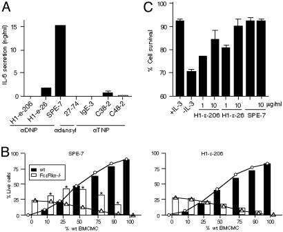
Fig. 1.
In vitro effects of HC and PC IgEs. (A) BMCMC were incubated with the indicated IgEs (5 μg/ml) for 8 h before measurement of IL-6 in culture supernatants. (B) Mixed cultures of WT and FcεRI α–/– BMCMC were incubated with 10 μg/ml SPE-7 or H1 DNP-ε-206 IgE for 3 days without IL-3 or other growth factors before flow cytometric analysis for FcεRI and annexin V. ○, Predicted survival values for WT cells; ▵, predicted survival values for FcεRI α–/– cells. These values are based on the assumption that each type of cell does not affect the survival of the other. Actual results are indicated by filled (WT) or open (FcεRI α–/–) bars. Asterisks indicate differences that are statistically significant (P < 0.05) from the predicted values. (C) WT BMCMC were incubated with or without IL-3, or with the indicated IgE without IL-3, for 3 days before flow cytometric analysis of cell survival.

