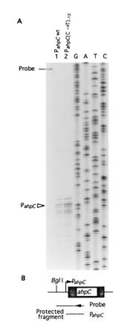
Figure 2.

S1 nuclease protection mapping of the ahpC mRNA 5′ end in M. bovis. (A) RNA was isolated from M. bovis bacillus Calmette–Guérin (BCG) ATCC35735 (lane 1) and M. bovis BCG ATCC35747 (lane 2). These strains have been examined for AhpC production (18) and displayed same relationships as shown in Fig. 1. Bar, location of the untreated probe. (B) Schematic representation of the probe and protected fragment in relationship to ahpC and its upstream region. Single-stranded S1 nuclease probe was generated using the same primer that served to generate DNA sequencing ladder (GATC). BglI site was the other end of the probe. Triangle (PahpC), ahpC mRNA 5′ end; wt, wild type; (C → T)−12, C → T transition at position −12 relative to the mRNA start site.
