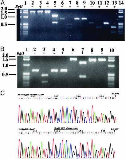
Fig. 3.
icSARS-CoV marker mutations. cDNAs spanning the different BglI junctions were amplified from WT and icSARS-CoV-infected cells. The fragments were purified, and a portion was digested with BglI and separated in 1.2% agarose gels. (A) icSARS-CoV (lanes 2, 3, 6, 7, 10, and 11) and WT SARS-CoV (lanes 4, 5, 8, 9, 12, and 13). Lanes 2–5, a 1,668-nt amplicon (nucleotide positions 1007–2675) digested with BglI (lanes 3 and 5); lanes 6–9, a 799-nt amplicon spanning the SARS-CoV B/C junction (positions 8381–9180) digested with BglI (lanes 7 and 9); lanes 10–13, a 544-nt amplicon (positions 11,721–12,265) spanning the SARS-CoV C/D junction digested with BglI (lanes 11 and 13). Lanes 1 and 14, a 1-kb ladder. (B) WT SARS (lanes 4, 5, 8, and 9); icSARS-CoV (lanes 2, 3, 6, and 7). Lanes 2–5, a 652-nt amplicon spanning the SARS-CoV D/E junction digested with BglI (lanes 3 and 5); lanes 6–9, a 1,594-nt amplicon (positions 23,665–25,259) spanning the SARS-CoV E/F junction digested with BglI (lanes 7 and 9). Lanes 1 and 10, a 1-kb ladder. (C) The 1,594-nt SARS E/F junction-containing amplicon was subcloned and sequenced, demonstrating the mutations introduced within the icSARS-CoV.

