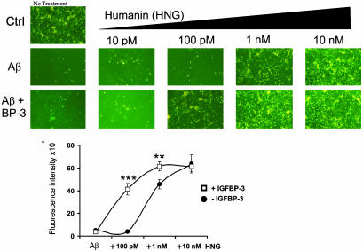
Fig. 5.
IGFBP-3 enhances HN protection against Aβ1–43 toxicity. Mouse cortical neurons were plated on poly-l-lysine-coated 96-well plates (5 × 10⁴ cells per well). Neurons were treated for 72 h with 25 μM Aβ1–43 with or without IGFBP-3 (10 nM) and with or without HN (10 pM to 10 nM). Representative photomicrographs are shown from calcein-stained cells at excitation 485 nm and emission 535 nm. Quantification of calcein fluorescence intensity is shown (Lower) (n = 3; **, P < 0.001; ***, P < 0.0001).

