
Figure 3.
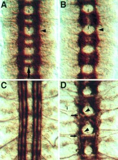
Phenotype of the kuz mutations. Each panel shows three segments of the CNS, and the large arrow in A marks the midline. (A) A wild-type stage 16 embryo stained with mAb BP102, which stains all CNS axon pathways. The arrowhead shows a longitudinal connective in the intercommissural region. The small downward arrow marks the anterior commissure, and the upward arrow indicates the posterior commissure, in one of the segments. (B) A kuz mutant stage 16 embryo stained with mAb BP102. Note the reduction in the longitudinal connectives (arrowhead) in contrast to the intense staining in the commissural region. The commissures (small arrows) are less distinct. (C) A wild-type stage 17 embryo stained with mAb 1D4, which recognizes the Fas II protein. At this stage Fas II is expressed on three parallel axon bundles on either side of the midline. (D) A kuz mutant stage 17 embryo stained with mAb 1D4. The three parallel bundles fail to form. Instead, most axons stall as large clumps in the commissural region (arrowheads). Only a small number of the Fas II-positive axons make it through the longitudinal connective (large arrows). In contrast to the CNS, the axons of the aCC neuron-pioneered intersegmental nerve (small arrow) extend large distances into the periphery, indicating that the CNS axon stalls are not due to a failure in the axon growth machinery.