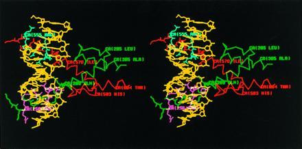
Figure 3.

Stereo diagram of the predicted structure of ato/da bHLH domain heterodimer–DNA complex by computer modeling. The sequence of the DNA-binding site in this model is TCAACAGCTGTTGA, containing an E box. DNA is colored in yellow, ato in green, and da in red. Those residues of ato and da predicted to contact DNA are colored in magenta and cyan, respectively. Only the side chains of the residues in the b domains are shown.
