Figure 4.

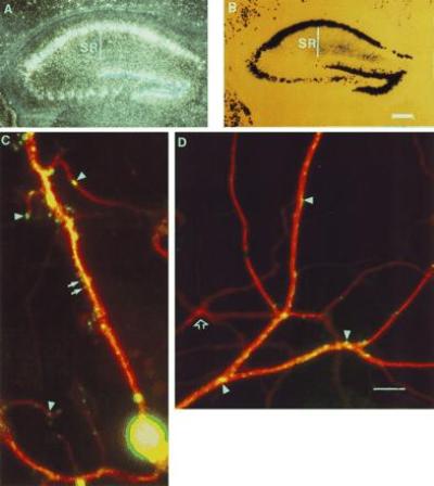
Differential expression of β-gal within dendrites. (A) In situ hybridization against the nls-lac-CMK mouse using a lacZ specific probe. (B) Histochemical detection of β-gal in the nls-lac-CMK mouse hippocampus (Bar = 300 μM.) (C) Immunofluorescent detection of MAP2 and β-gal expression in an nls-lac-CMK neuron in culture. The MAP2 antibody specifically labels microtubules along the dendritic shaft. MAP2 labeling is indicated in red. β-gal labeling is shown in green. Arrows denote β-gal in presumptive dendritic spines. Arrowheads indicate areas of punctate β-gal staining along the dendrite. (D) Expression of β-gal in a distal portion of the dendritic arbor. Arrowheads denote areas of punctate β-gal staining. Open arrow shows a dendrite arising from a neuron, which did not express the nls-lac-CMK transgene (Bar = 10 μM.)
