
Figure 5.
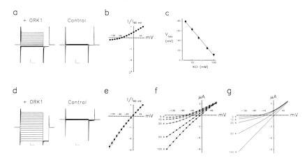
ORK1 currents in X. laevis
oocytes. (a) ORK1 currents in physiological levels of
[K]o are outwardly rectifying. Currents were assessed in
oocytes injected with 1 ng of cRNA (+ ORK1) or water (control) by
two-electrode voltage clamp under constant perfusion with 5 mM KCl
solution. Oocytes were pulsed from −150 to 60 mV in 15-mV steps for 75
ms followed by a 15-ms step to −150 mV before returning to the holding
potential of −80 mV; a 1-s interpulse interval was employed. Currents
are displayed without leak subtraction. Scale bars represent 2 μA and
15 ms. (b) ORK1 current–voltage relation in 5 mM KCl
solution at 10 ms into the test pulse normalized to current at 60 mV by
the protocol in a (mean ± SEM,
n = 4 cells). (c) ORK1 currents are
K+ selective. The reversal potential of currents was
studied with 5, 10, 20, 50, or 100 mM KCl solutions by the protocol in
a (mean ± SEM, n = 4 cells).
Linear regression gives a shift of 55 ± 2 mV per 10-fold change
in KCl concentration. (d) ORK1 currents flow inward at
hyperpolarized voltages under constant perfusion with 100 mM KCl
solution; protocol and scale bars as in a.
(e) ORK1 current–voltage relation in 100 mM KCl
solution as in b (mean ± SEM,
n = 4 cells). (f) ORK1
current–voltage relation for one oocyte studied in 5, 10, 20, 50, and
100 mM KCl solutions as in b. (g)
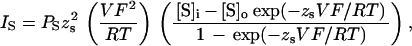
Theoretical current–voltage relations under the conditions used to
study ORK1 in e according to Goldman (17) and Hodgkin
and Katz (18):
where Ps is the permeability of
K+, [S] refers to K+ concentration, and
z, V, F, R, and
T have their usual meanings, and assuming an internal
K+ concentration of 90 mM, as reported previously (35).
 |