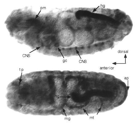
Figure 4.

Whole-mount in situ localization of ine transcripts in late-stage embryos. (Upper) Lateral view. (Lower) Dorsal view. Strong staining can be seen in the posterior hindgut (hg), anal plate (ap), Malpighian tubules (mt), midgut (mg), tracheal pits (tp), pharyngeal muscle region (pm), and a subset of segmentally repeating cells in the central nervous system. Additional small patches of specific staining in the head were difficult to identify. Embryo preparation, fixation, hybridization, and staining were conducted as described (31). The probe was prepared from antisense RNA transcribed from the ine cloned cDNA using SP6 RNA polymerase (Boehringer Mannheim). Probe of sense RNA prepared from T7 RNA polymerase (Boehringer Mannheim) was ulilized as a control. The probe contained 200–300 bases from the 5′ end of the ine cDNA. The color reaction was allowed to proceed for 10 h.
