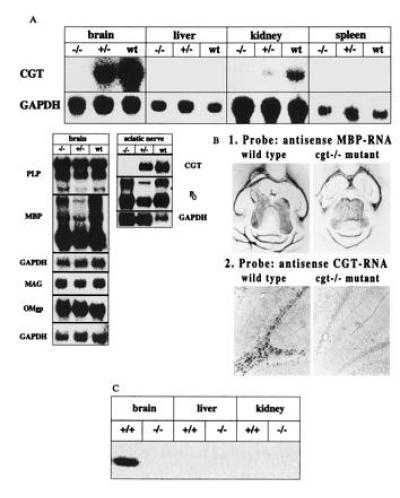
Figure 2.

Analysis of cgt expression and relevant myelin-associated genes of oligodendrocyte by Northern blot, in situ hybridization and enzyme assay. C57BL/6 mice were used as control mice in all comparative experiments. (A) Northern blot analysis of CGT, PLP, MBP, MAG, OMgp, and P0 RNA of brain, liver, kidney, spleen, and sciatic nerve. Approximate sizes of transcripts: CGT, 3.2 kb; PLP, 1.6, 2.4 and 3.2 kb; MBP, 2.1 kb; MAG, 4 kb; OMgp, 1.8 kb; Po, 2.0 kb; glyceraldehyde-phosphate dehydrogenase, 1.2 kb. (B) Comparative in situ hybridization analysis of CGT and MBP expression in wt and homozygous cgt−/− mouse brain. (C) CGT activity of brain, liver and kidney of age matched p18-20 wt and homozygous mutant mice. Enzymatic activity was determined as described (17) with synthetic d-2-hydroxyhexanoylsphingosine and UDP[14C]galactose (Amersham) as substrates and microsomal protein of brain, liver, and kidney from p18 wt and cgt−/− mice. The reaction product, radioactive [14C]galactosyl ceramide, was detected in the lipid extract after separation by TLC as described and recorded with the phosphoimager. Only brain microsomes of wt mouse brain showed significant enzymatic activity, that of kidney was to weak to be detected by autoradiography.
