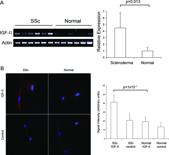
Figure 2.
Steady-state IGF-II mRNA (A) and protein levels (B) are increased in SSc lung fibroblasts. A: RT-PCR analysis of total IGF-II mRNA levels in primary lung fibroblasts shows significantly increased expression in SSc versus normal fibroblasts (n = 6, 4.5 ± 2.3 versus 1.0 ± 0.5, respectively; P = 0.013). Relative expression was determined by scanning densitometry of PCR products normalized to β-actin mRNA levels. B: IGF-II protein was detected by immunofluorescence using polyclonal anti-IGF-II antibody and compared to goat isotype control antibody. Signal intensity was quantified with Metamorph imaging software (Molecular Devices) and shown in arbitrary units. SSc lung fibroblasts show a twofold increase in IGF-II protein compared to normal lung fibroblasts (n = 4, P = 1 × 10⁻⁷). Values and bars represent mean and SD, respectively. Original magnifications, ×400.

