Figure 2.
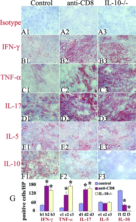
Effect of CD8 depletion and IL-10 deficiency on protein expression of pro- and anti-inflammatory cytokines. Isotype control staining (A1–A3) was always negative. Representative thyroids in each group at day 20 expressed the proinflammatory cytokines IFN-γ (B1–B3), TNF-α (C1–C3), and IL-17 (D1–D3) and anti-inflammatory cytokines IL-5 (E1–E3) and IL-10 (F1–F3). The staining intensity for proinflammatory cytokines was stronger in thyroids of CD8-depleted WT and IL-10−/− recipients and staining intensity for IL-10 was weaker or undetectable compared to WT controls. The staining intensity for IL-5 was similar for all groups. Cytokine-positive cells (red) in five to six randomly selected high-power fields of three representative thyroids in each group were manually counted using an enlarged image. The results are summarized in G (bars b1–f3 correspond to B1–F3). A significant difference between CD8-depleted WT and IL-10−/− recipients versus WT controls is indicated by the asterisk (P < 0.05). Results are representative areas on slides from at least three individual mice examined per group with comparable G-EAT severity scores (4 to 5+). Original magnifications, ×400.

