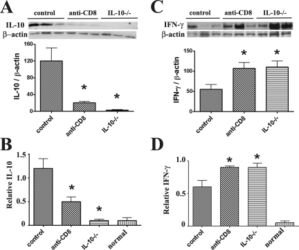
Figure 3.
Quantitative determination of the effect of CD8 depletion and IL-10 deficiency on IL-10 and IFN-γ protein and mRNA expression At day 20, IL-10 protein was undetectable in thyroids of IL-10−/− recipients of IL-10−/− splenocytes (A) and IL-10 was lower in thyroids of CD8-depleted WT recipients compared to WT controls. Real-time quantitative PCR indicated that IL-10 mRNA was undetectable in thyroids of IL-10−/− recipients of IL-10−/− splenocytes (B) and IL-10 mRNA was lower in thyroids of CD8-depleted recipients compared to WT controls. IFN-γ protein (C) and mRNA (D) expression was higher in thyroids of CD8-depleted WT or IL-10−/− recipients compared to WT controls. For Western blot, 30 μg of protein from thyroids of each group (three individuals) was loaded in each lane and results are shown at the top in A and C. Results are expressed as the mean ratio of densitometric U/β-actin ± SEM (×100), and are representative of two independent experiments. For real-time PCR, bars are means of data for thyroids of four to five individual mice ± SEM. Results are expressed as the mean relative ratio to HPRT. A significant difference between CD8-depleted WT and IL-10−/− recipients versus WT controls is indicated by the asterisk (P < 0.05).

