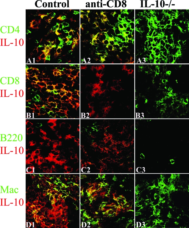
Figure 4.
Cell distribution of IL-10 protein in thyroids of CD8-depleted recipients and WT controls. Dual-color immunofluorescence and confocal laser-scanning microscopy on thyroid frozen sections 20 days after cell transfer. CD4+ and CD8+ T cells, B cells, and macrophages were identified by cell surface expression of CD4, CD8, B220, or F4/80 (A1–D3, green), and IL-10-producing cells were identified by IL-10 staining (red). IL-10 was undetectable in thyroids of IL-10−/− recipients (A3–D3, red) and few CD8+ T cells were detected in CD8-depleted WT recipients (B2, green). IL-10 was produced by CD4+ T cells (A1, yellow, overlay), CD8+ T cells (B1, yellow, overlay), and macrophages (D1, yellow, overlay) in thyroids of WT controls. After depletion of CD8+ T cells, most IL-10 was produced by CD4+ T cells (A2, yellow, overlay) and macrophages (D2, yellow, overlay). No IL-10+ B cells were detected (C1–C3). CD8+ T cells (B1, yellow plus green) outnumbered CD4+ T cells (A1, yellow plus green) in WT controls, whereas CD4+ T cells outnumbered CD8+ T cells in CD8-depleted WT or IL-10−/− recipients (A1–A3 versus B1–B3). Shown are representative areas on slides of thyroids of at least three individual mice per group with comparable 4 to 5+ G-EAT severity scores. Original magnifications, ×800.

