Figure 6.
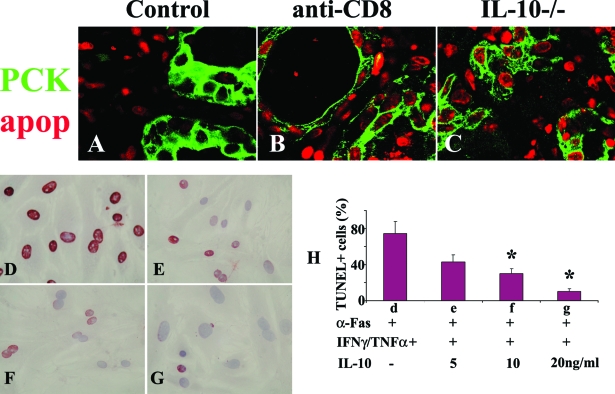
IL-10 protects TECs from Fas-mediated apoptosis in vitro and in vivo. A–C: The distribution pattern of apoptotic cells in each group was determined by a fluorescence apoptosis kit. TECs were identified by pan-cytokeratin (PCK, green) and apoptosis was detected as red nuclear staining. Most apoptotic cells (red) were inflammatory cells in WT controls (A) and most were TECs in thyroids of CD8-depleted WT (B) and IL-10−/− recipients (C). Shown are representative areas on slides of thyroids of at least three individual mice per group with comparable G-EAT severity scores. Sixty to seventy percent confluent cultured TECs were pretreated with IFN-γ/TNF-α for 4 days in the presence (5 to 20 ng/ml) or absence of IL-10 and stimulated with anti-Fas for 20 hours. Apoptosis was detected by TUNEL staining. TUNEL+ cells (D–G, red) in five to six randomly selected high-power fields of three individual mice per group were manually counted and summarized in H (bars d–g correspond to D–G). In the absence of IL-10, TECs cultured with cytokines and anti-Fas underwent extensive Fas-mediated apoptosis (74 ± 13% TUNEL+ cells), whereas the percentage of TUNEL+ cells decreased in the presence of IL-10 in a dose-dependent manner. A significant difference in the percentage of TUNEL+ cells in the presence of different concentrations of IL-10 compared to no IL-10 is indicated by the asterisk (P < 0.05). Original magnifications: ×800 (A–C); ×400 (D–G).

