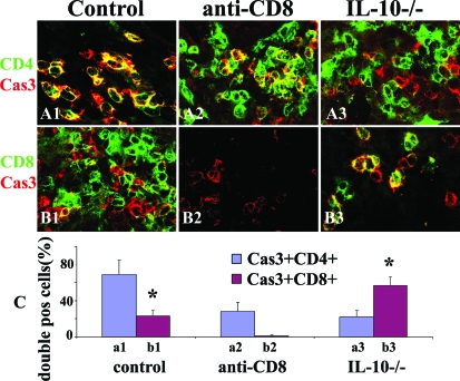
Figure 7.
CD8+ T cells in thyroids of mice with G-EAT are protected from apoptosis by IL-10. Confocal staining of CD4 and CD8 on frozen sections with active caspase-3 at day 20. Active caspase-3 (red, A1–B3) is positive in both CD4+ (green, A1–A3) and CD8+ (green, B1–B3) T cells (yellow, overlay). Apoptotic (active caspase-3+) inflammatory cells in controls are mainly CD4+ T cells (A1, yellow, overlay), whereas most apoptotic cells in IL-10−/− recipients of IL-10−/− donor cells are CD8+ T cells (B3, yellow, overlay). Active caspase-3+CD4+ or active caspase-3+CD8+ cells were manually counted in five to six randomly selected high-power fields and expressed as the percentage of total CD4+ or CD8+ T cells and summarized in C. Shown are representative areas on slides of thyroids of at least three individual mice per group with comparable 4 to 5+ G-EAT severity scores. A significant difference between the percentage of active caspase-3+CD4+ and active caspase-3+CD8+ cells in WT controls or in IL-10−/− group is indicated by the asterisk (P < 0.05). Original magnifications, ×800.

