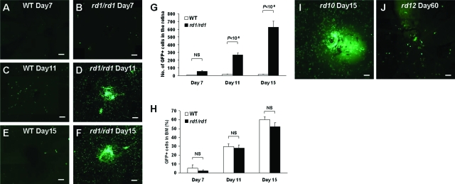
Figure 1.
BM-derived cells were recruited into the degenerating retina. A–F: Retinal flatmount after BMT with GFP donor mice. Seven days after BMT in wild-type (WT) mice (A) and rd1/rd1 mice (B). Eleven days after BMT in WT mice (C) and rd1/rd1 mice (D). Fifteen days after BMT in WT mice (E) and rd1/rd1 mice (F). G: GFP+ cell counts in the retina of WT mice and rd1/rd1 mice (n = 6). H: GFP+ cell percentage of BM in WT mice and rd1/rd1 mice (n = 6). NS, not significant. Retinal flatmount after GFP labeling and BMT in other degeneration models, rd10 (I) and rd12 (J). BMT was performed at P15 in rd10, and at P30 in rd12. The images were collected 15 days after BMT in rd10 and 60 days after BMT in rd12. Scale bars = 100 μm.

