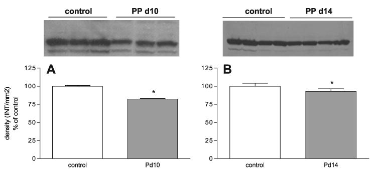
Fig. 5.

The expression of Cyp27a1 protein during PP days 10 and 14. Western blot analysis of Cyp27a1 protein expression in control and PP day 10 (A) and 14 (B) rats. Densitometric analysis of Cyp27a1 Western blots (n = 3–4 animals). Each bar represents the mean ± SD of Cyp27a1 density (INT/mm2). *P < 0.05 vs. control rats as determined by Student's t-test. For Western blot analysis, Ponceau red staining was performed for each membrane to ensure equal loading in each lane.
