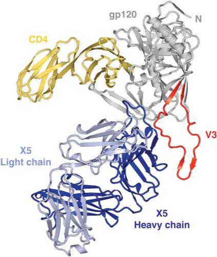
Fig. 1.

Structure of an HIV-1 gp120 core with V3. The crystal structure of core gp120 (gray) with an intact V3 (red) is shown bound to the membrane-distal two domains of the CD4 receptor (yellow) and the Fab portion of the ×5 antibody (dark and light blue). In this orientation, the viral membrane would be positioned toward the top of the page and the target cell toward the bottom.
