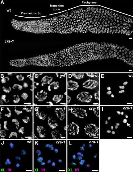
Figure 2. Chromosome Morphogenesis and Nuclear Organization During Meiotic Prophase Are Altered in cra-1 Mutants.
(A) Low magnification images of DAPI-stained nuclei of whole-mount gonads from age-matched wild type and cra-1(tm2144) adult hermaphrodites. Progression throughout early- and mid-prophase is observed from left to right. (B–I) High magnification images of DAPI-stained nuclei taken from the transition zone (B, F), early pachytene (C, G), late pachytene (D, H) and diakinesis (E, I) of wild type (B–E) and cra-1(tm2144) (F–I) depicting the defects observed in chromosome morphogenesis and nuclear organization. Arrow indicates a bivalent in the cra-1(tm2144) diakinesis oocyte in (I). (J–L) High magnification images of diakinesis oocytes stained with DAPI (blue) and hybridized with FISH probes targeting the left end of the X chromosome (green) and the right end of chromosome I (red) in wild type (J) and cra-1(tm2144) mutants (K–L). Bars, 5 µm (A) and 2 µm (B–L).

