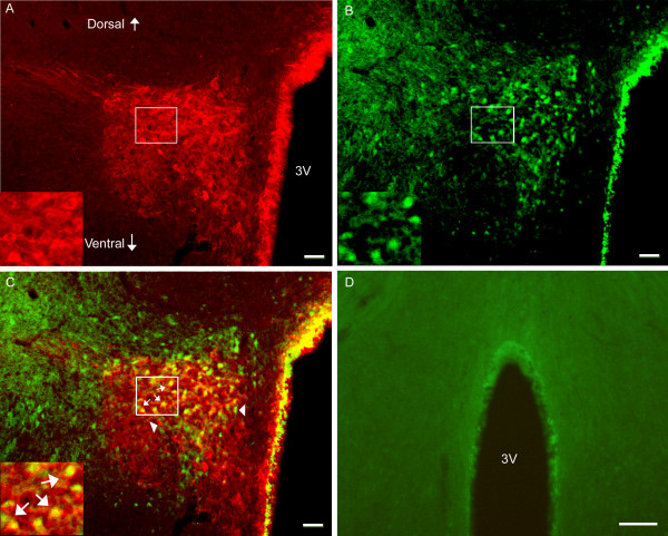
Figure 6.
Photomicrographs showing co-localization of CRH and p-NR1 in the PVN. A-C: Sections from EA-treated naive rats were double-labeled with anti-CRH (red) and anti-p-NR1 (green). A: CRH-immunoreactive neurons in the PVN. B: p-NRI-immunoreactive neurons in the PVN. C: Merged graphs of A and B. Small arrows indicate examples of double-labeled CRH/p-NR1 neuron (yellow); Arrowheads point to single-labeled CRH and p-NR1. The insets in A, B and C are higher magnification of the square areas in A, B and C, respectively. D: Sections from untreated naive rats were signle-labeled with anti-p-NR1 and showed no labeling of P-NR1. Scale bars represent 50 μm in A, B, C, and 250 μm in D.

