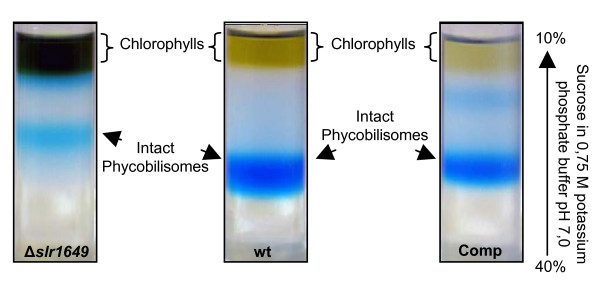
Figure 4.
Isolation of intact phycobilisomes. Phycobilisomes were isolated as described in Material & Methods. After 16 h centrifugation the phycobilisomes became visible as clear blue bands in the gradient. The upper layer contained chlorophylls. The phycobilisomes of Δslr1649 cells had a diminished migration compared to the wild type ones, whereas the phycobilisomes from the complemented strain (Comp) had a migration equivalent to the wild type.

