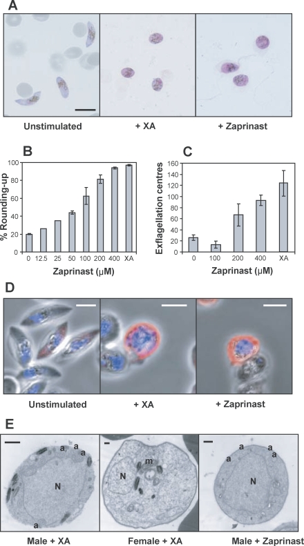
Figure 1. Zaprinast Can Stimulate Rounding Up and Exflagellation of Mature Gametocytes in the Absence of XA.
Zaprinast was added to mature gametocytes to assess the effects on gametogenesis compared to XA. The concentration of XA used was 20 μM, and the concentration of zaprinast was 400 μM, unless stated otherwise.
(A) Micrographs of Giemsa-stained Stage V gametocytes prior to stimulation of gametogenesis (left panel) and after addition of XA (centre panel) or zaprinast (right panel). The scale bar indicates 10 μm.
(B) Increasing concentrations of zaprinast were added to stimulate gametogenesis, and cells were scored as either round or crescent-shaped, and plotted as a percentage rounded-up. Results are based on triplicate counts of a representative experiment from the same flask of gametocytes on a single day (except for 12.5 and 25 μM, which are based on a single count only). Error bars indicate the standard error of the mean (± SEM). The experiment was carried out twice with very similar results.
(C) The number of centres of exflagellation per 10,000 gametocytes was scored following addition of increasing concentrations of zaprinast. Results are based on triplicate counts of a representative experiment from the same flask of gametocytes on a single day. Error bars indicate mean ± SEM. The experiment was carried out twice with very similar results.
(D) Merged confocal images of Giemsa-stained stage V gametocytes prior to stimulation of gametogenesis (left panel) and after addition of XA (centre panel) or zaprinast (right panel). Blue indicates DAPI-stained nuclei and red the anti-α tubulin antibody (Tat1) staining of cells. The scale bars indicate 5 μm.
(E) Transmission electron micrographs of male or female gametocytes after stimulation of gametogenesis with XA (left and centre panels) or zaprinast (right panel). a, axonemes; m, mitochondrion; N, nucleus. Scale bars indicate 0.5 μm.

