Fig. 2.
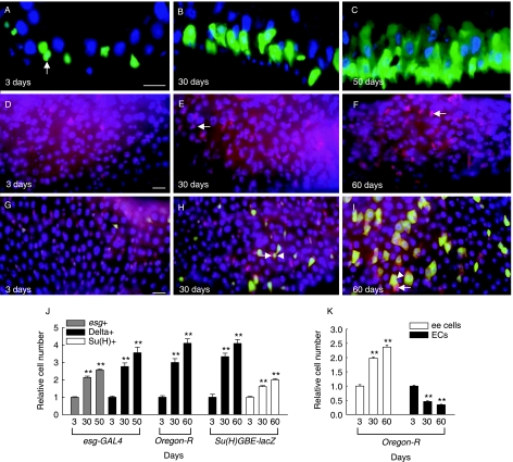
Age-related changes in the number of ISCs/and progenitor cells within the adult midgut. (A–C) Age-related change in the number of esg-positive cells (ISCs/or enteroblasts) in the adult midgut. Cross-sections of the posterior midguts of 3-, 30- and 50-day-old esg-GAL4,UAS-GFP/CyO flies were conducted with Cryostat and labeled with anti-GFP. Arrow indicates GFP-expressing cells under the control of esg-GAL4. Overlay (DAPI, blue; anti-GFP, green). (D–F) Age-related change in the number of Delta-positive cells (ISCs). The posterior midguts of 3-, 30- and 60-day-old wild-type flies were stained with anti-Delta (arrow). Overlay (DAPI, blue; anti-Delta, red). (G–I) Age-related change in the number of Su(H)-positive cells (enteroblasts). The posterior midguts of 3-, 30- and 60-day-old Su(H)GBE-lacZ flies were stained with anti-Delta (arrow) and anti-β-gal (arrowhead). Overlay (DAPI, blue; anti-β-gal, green; anti-Delta, red). All scale bar, 20 µm. (J) Graph showing age-related the relative numbers of esg-, Delta- or Su(H)-positive cells detected in the posterior midguts of esg-GAL4,UAS-GFP/CyO, wild-type and Su(H)GBE-lacZ flies. The cell numbers detected in the posterior midgut of 3-day-old flies were set at 1. For quantitative method of GFP-, Delta- and β-gal-expression in the posterior midgut, images were processed in Adobe Photoshop and then the cell number was counted in 0.06 × 0.02 cm area of the posterior midgut. The results are expressed as the mean ± SE values of 19–69 adults. (K) Graph showing age-related the relative numbers of enteroendocrine (ee) cells and enterocytes (ECs) with large nuclei detected in the posterior midgut of wild-type flies. The cell numbers detected in the posterior midgut of 3-day-old flies were set at 1. The midguts of 3-, 30- and 60-day-old wild-type flies were stained with anti-Prospero or DAPI, after which the numbers of enteroendocrine cells or enterocytes with large nuclei were assessed. For quantitative analysis, the images were processed in Adobe Photoshop and the number of enteroendocrine cells or enterocytes with large nuclei was counted in 0.12 × 0.02 cm or 0.06 × 0.02 cm area of the posterior midgut, respectively. The results are expressed as the mean ± SE values of 28–50 adults. All two asterisks represent P < 0.001 as compared to each 3-day-old flies.

