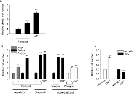
Fig. 3.
Oxidative stress-induced changes in the number of ISCs and progenitor cells within the adult midgut. (A) Effects of paraquat treatment and the Catn1 allele on the number of phospho-histone H3-positive cells. The midguts of 5-day-old wild-type flies exposed to 10 mm paraquat in 1% sucrose or 1% sucrose media as controls for 16 h, and 5-day-old Catn1 mutant or wild-type flies as controls were labeled with anti-phospho-histone H3. Graph shows the relative number of phospho-histone H3-positive cells per midgut. The number of phospho-histone H3-positive cells observed per midgut of wild-type flies was set at 1. Results are expressed as the mean ± SE values of 39–52 adults. (B) Effects of paraquat treatment and the Catn1 allele on the number of ISCs and progenitor cells in the adult midgut. The midguts of 5-day-old esg-GAL4,UAS-GFP/CyO, wild-type and Su(H)GBE-lacZ flies exposed to 10 mm paraquat in 1% sucrose or 1% sucrose media as control for 16 h, 5-day-old wild-type, Catn1 mutant, Su(H)GBE-lacZ/+ and Su(H)GBE-lacZ/Catn1 flies were labeled with anti-GFP, anti-Delta and anti-β-gal. Graph shows the relative numbers of esg-, Delta- or Su(H)-positive cells in the posterior midgut. The cell numbers detected in the posterior midgut of paraquat-untreated flies, wild-type and Su(H)GBE-lacZ/+ flies were set at 1, respectively. For quantitative method of GFP-, Delta- and β-gal-expression in the posterior midgut, images were processed in Adobe Photoshop and then the cell number was counted in 0.06 × 0.02 cm area of the posterior midgut. The results are expressed as the mean ± SE values of 16–35 adults. (C) Effects of Catn1 allele on the number of enteroendocrine cells and enterocytes with large nuclei in the adult midgut. Graph shows the relative numbers of enteroendocrine (ee) cells and enterocytes (ECs) in the adult posterior midguts of Catn1 mutant flies as compared to wild-type flies. The cell numbers detected in the posterior midgut of wild-type flies were set at 1. The midguts of 5-day-old wild-type and Catn1 mutant flies were stained with anti-Prospero or DAPI, after which the numbers of enteroendocrine cells or enterocytes with large nuclei were assessed. For quantitative analysis, the images were processed in Adobe Photoshop and then the number of enteroendocrine cells or enterocytes with large nuclei was counted in 0.12 × 0.02 cm or 0.06 × 0.02 cm area of the posterior midgut, respectively. The results are expressed as the mean ± SE values of 31–40 adults. All two asterisks represent P < 0.001 as compared to each control.

