
fig. 2.
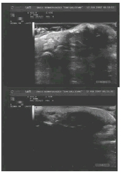
Real-time US using 13-MHZ linear probe; sonographic longitudinal section of the distal interphalangeal joint showed well-defined thickness of the nail matrix and nail bed, with slight eye catching signal at power Doppler, together with superficial soft tissue thickening at the distal phalanx.