
Figure 1.
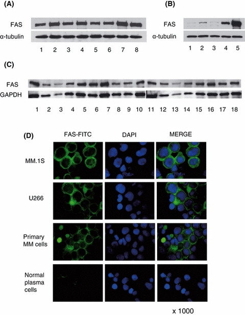
FAS expression in various cells. Cell lysates (20 μg) of MM cell lines (A), normal cells and MM cells (B), and patient cells (B; lane 4, C) were immunoblotted with anti‐FAS antibody. (A) FAS expression was detected in all MM cell lines: lane 1, U266; lane 2, MM.1S; lane 3, MM.1R; lane 4, RPMI8226; lane 5, RPMI Dox40; lane 6, RPMI LR5; lane 7, OPM1; and lane 8, OPM2. (B) FAS expression level was compared in plasma cells and MM cells (lane 1–3, normal plasma cells; lane 4, primary MM cells; lane 5, MM.1S). FAS protein was more highly expressed in MM.1S and primary MM cells than in plasma cells. (C) FAS protein was expressed in all (18/18) primary MM cells. (D) FAS expression in MM cell lines, primary MM cells and normal plasma cells was analysed by immunocytochemistry. FITC‐labeled FAS, nuclear staining by DAPI, and combined staining (Merge) were evaluated by fluorescence microscopy (×1000). Green and blue signal show FAS‐FITC and DAPI respectively. FAS protein in MM cells is most abundant in the cytoplasm with only weak non‐specific of nuclear membrane staining.