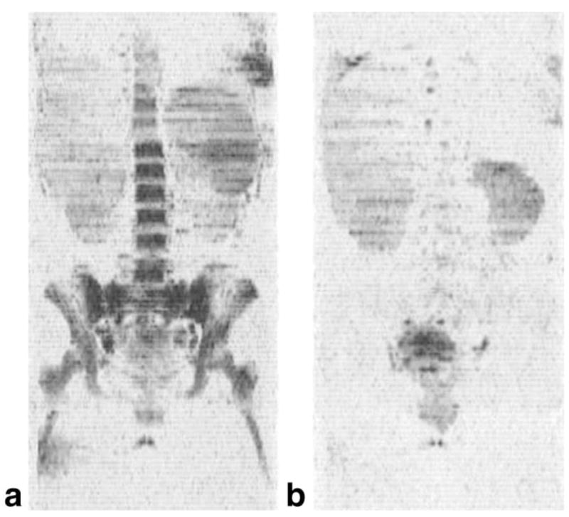
FIG. 2.
The techniques applied to a subject with acute myelogenous leukemia: (a) coronal image from a baseline study obtained just prior to initiation of myelosuppressive therapy, (b) a second examination obtained 20 days later after the patient had achieved a remission. The bone marrow was intact at baseline with a water fraction of 0.99 ± 0.06 and a T2 of water equal to 61.7 ± 1.8 msec, which is consistent with the presence of leukemia. Twenty days after the start of chemotherapy, the water signal in the bone marrow on the T2- and diffusion-weighted images is virtually absent. The patient had achieved a complete remission at this time, with only 2% myeloblasts on the differential blood count.

