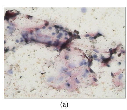
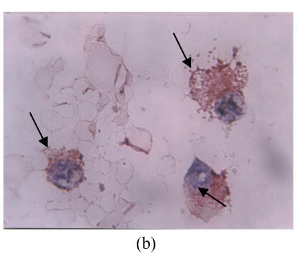
Fig. 1.
Immunohistochemical expression of FAS in bone marrow samples from multiple myeloma patients. (a) Low magnification micrograph (200×) showing FAS expression in a bone marrow smear of a sample obtained from one MM patient; (b) Higher magnification (1000×) of the same sample illustrating the cytoplasmic localization of FAS in the myeloma plasma cells. Cells with arrows are the FAS positive; (c) Low magnification micrograph (200×) of FAS expression in one negative sample obtained from one MM patient


