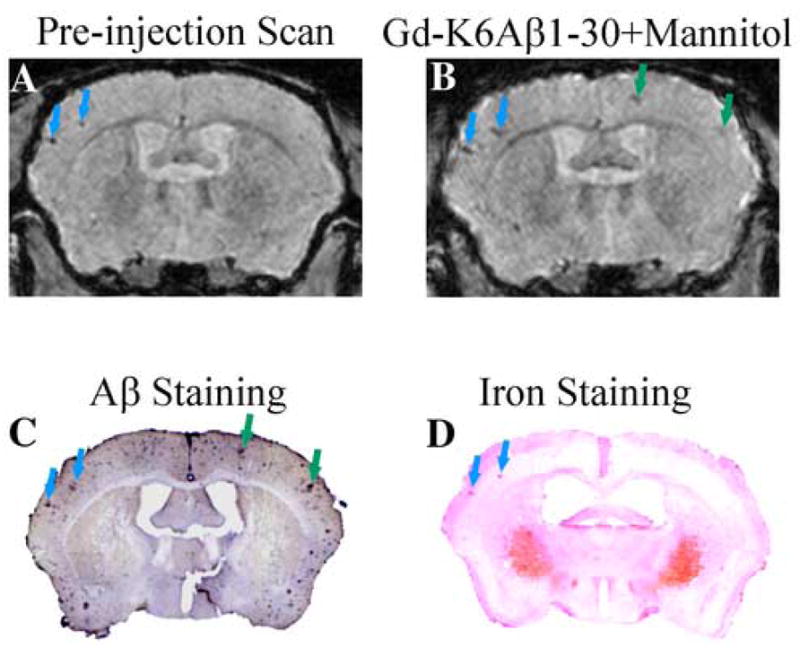
Figure 3.

Detection of Aβ plaques without contrast agent correlates with plaques detected by Perl’s iron stain. (A) A few large plaques are detected in the parietal cortex in the pre-ligand injection scan of a 20 month old Tg2576 mouse (blue arrows). Following the intracarotid injection of a contrast agent (Gd-DTPA-K6Aβ1–30), the intensity of these plaques is enhanced and additional plaques are detected (green arrows). These plaques co-register with Aβ deposits on tissue section (C), and the plaques detected without a contrast agent stain for iron as depicted in D. At this magnification, the Perls’ stained iron deposits (blue) appear pink because of the nuclear fast red counterstain used for nuclei and cytoplasm.
