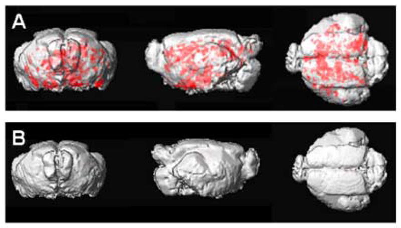
Figure 4. 3D rendering of cortical Aβ plaques.

(A) A three-dimensional rendering of the transgenic mouse template showing the distribution of amyloid deposits as depicted in red, in the MRI images following Gd-DTPA-K6Aβ1–30 injection.
(B) The same three-dimensional rendering of the wild-type mice template shows no amyloid deposits in the MRI images following Gd-DTPA-K6Aβ1–30 injection in the group of wild-type mice.
