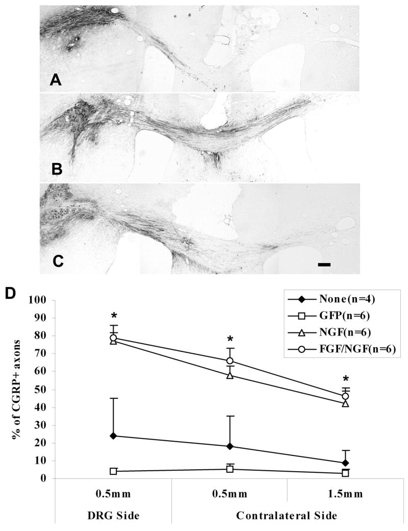
Figure 4.

CGRP+ axons have grown around the lesion into the contralateral corpus callosum by 2 weeks post-transplant. A) Representative section from an animal with GFP expression along the pathway. Very few CGRP+ fibers grew around the lesion into the contralateral corpus callosum. B and C) Representative sections from the animals with either NGF (B) or FGF/NGF (C) expression along the pathway. More CGRP+ fibers grew around the lesion into the contralateral corpus callosum. D) Percentage of CGRP+ fibers that grew around the lesion of the corpus callosum. This shows in a similar pattern as in Figure 3, with a higher percentage of CGRP+ axons that grew around the lesion into the contralateral side in both NGF and FGF/NGF pathways. *p<0.001, FGF/NGF, NGF vs GFP, p<0.01, FGF/NGF, NGF vs None (no pathway); p>0.05, FGF/NGF vs NGF; GFP vs None (no pathway). Data = Mean ± SEM. Scale bar = 200μm.
