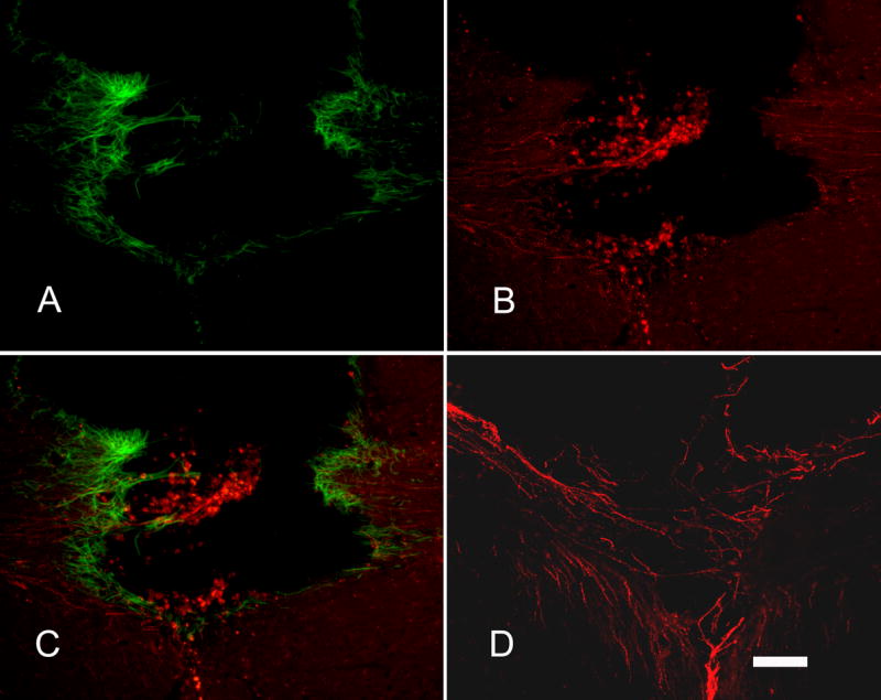
Figure 6.

CGRP+ axons grew into the lesion area where reactive astrocytes were present around the lesion. There was no difference in GFAP staining between groups. A) Reactive astrocytes stained with GFAP (green) antibody accumulated around the lesion site. B) CGRP+ axons (red) from DRG transplants grew into the lesion area; some grew into the opposite corpus callosum. C) Merged image of A and B to demonstrate the relationship between axon growth and glial scar around the lesion site. D) Some CGRP+ axons (red) are seen bridging the lesion site to grow into the opposite corpus callosum. Sections were from NGF pathway group in A, B, C. Section was from FGF/NGF pathway group in D. Scale bar = 100μm.
