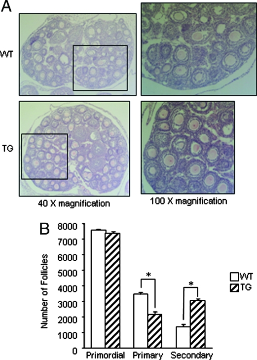
Figure 2.
Follicle development in wild-type (WT) and transgenic (TG) ovaries. A, Histology of ovaries from 25-d-old wild-type and transgenic mice. Ovaries were photographed at ×40 (left panels) magnification. The boxed areas are shown at ×100 magnification (right panels). B, Numbers of primordial, primary, and secondary follicles were determined in wild-type and transgenic ovaries at 25 d of age. Ovaries were serially sectioned and every fifth section was counted, and total follicle numbers were determined. These data represent the mean ± sem of combined results from analysis of four mice per age and genotype (*, P ≤ 0.05).

