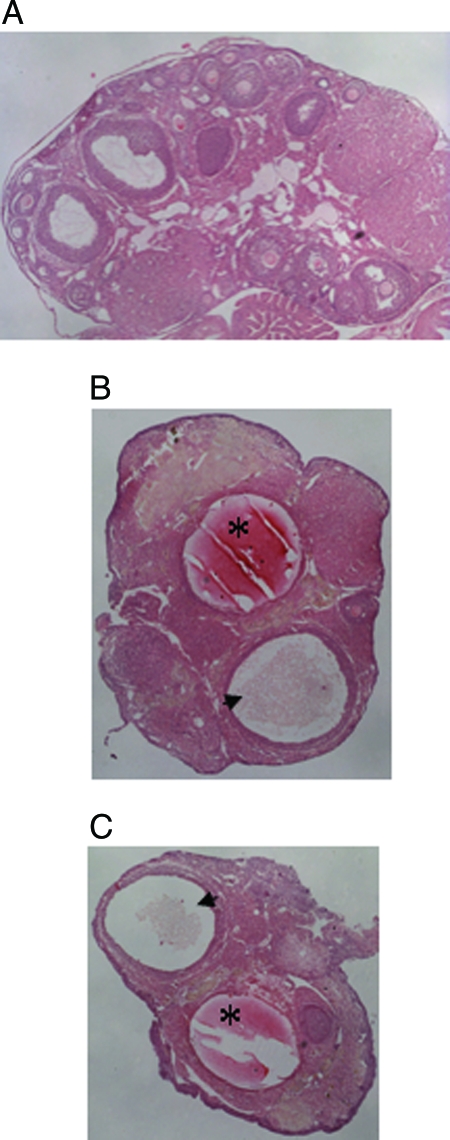
Figure 9.
Histological analysis of transgenic ovaries. Ovarian sections were stained with hematoxylin and eosin and photographed at ×40 magnification. A, Normal ovary from a cycling mouse exhibiting follicles of various developmental stages and corpora lutea. B and C, Ovaries from transgenic mice that had entered constant diestrus, exhibiting hemorrhagic (asterisk) and fluid-filled (arrowhead) cysts and a lack of developing follicles.

