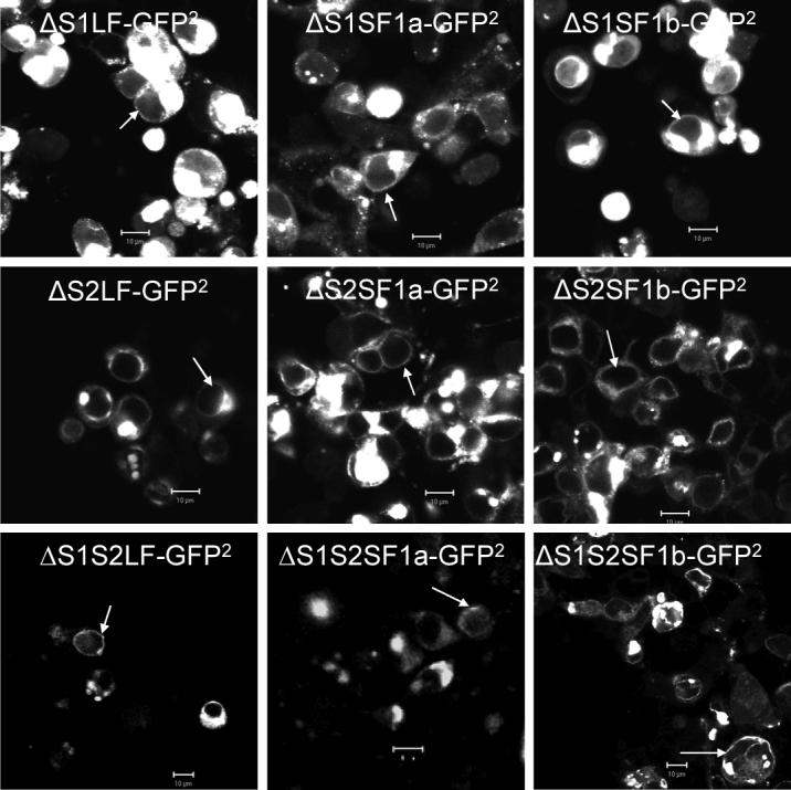
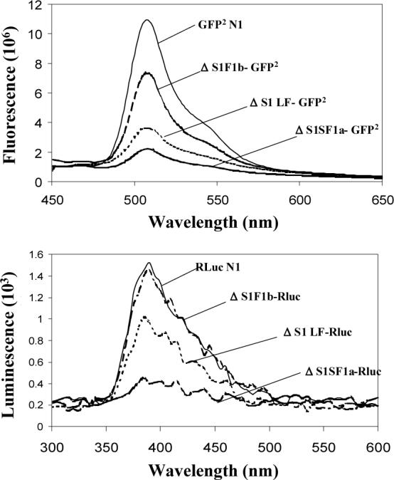
Fig 2. Confocal images demonstrating expression, localization (A) and spectral properties (B) of the GFP2 and/or Rluc-tagged deleted human PRLRs.


HEK 293 cells transiently transfected with plasmids containing cDNA coding for the receptors tagged with GFP2 were examined by confocal microscopy after 48h. bar,10μm; white arrowhead, localization of the tagged receptors to the region of the plasma membrane. For spectral scanning, GFP2 was excited at 405nm. Bioluminescence generated by Rluc was detected in the presence of substrate, DeepBlueC (5 μM), with blockade of the external excitation light source. Only the spectra for the ΔS1 versions are shown by way of example, but all spectra were unaltered by attachment to the various receptors. GFP2N1 is the GFP2 expressing vector without any receptor, and RLuc1N1 is the luciferase expressing vector without receptor. The relative expression of each receptor can be appreciated by comparing the amount of fluorescence or luciferase activity. Relative expression levels were the same for the intact and deleted versions of the receptor.
