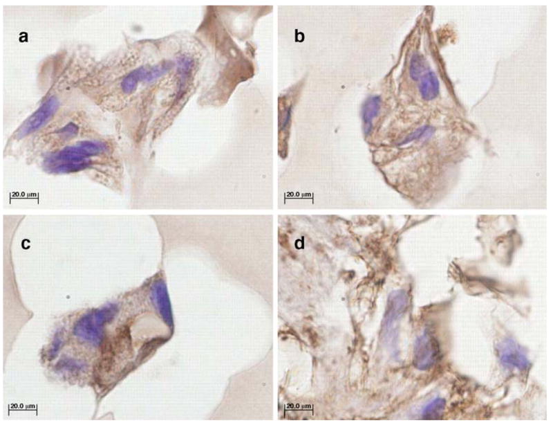
Fig. 5.

Immunohistochemical staining of paraffin-embedded sections -TGF-β3 (a and c) and + TGF-β3 (b and d) hMSC/SELP-47 K constructs with antibodies for type I collagen (a and b) and type II collagen (c and d) after 28 days of culture. Controls showed no staining for antigen (not shown). Images were originally acquired at 100X magnification. Scale bars=20 μm. Dark brown staining is positive for the collagen.
